resources


resources

Tuesday, February 17, 2009
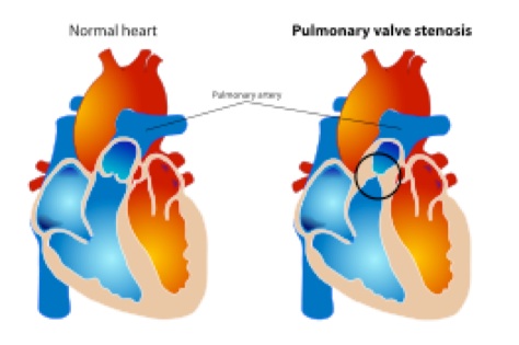
Pulmonary valve stenosis is a condition in which the flow of blood from the heart (right ventricle, or lower chamber) is blocked at the valve that separates the heart from the pulmonary artery (pulmonic valve). This narrowing is usually present at birth (congenital).
Causes
Pulmonary valve stenosis is most often caused by a problem that occurs when the unborn baby (fetus) is developing. The cause is unknown, but genetics may play a role.
Narrowing that occurs in the pulmonary valve is called pulmonary valve stenosis. Narrowing that occurs below the pulmonary valve is called subvalvar pulmonary stenosis. Another form of the condition, supravalvar pulmonary stenosis, is when narrowing occurs above the main pulmonary valve.
The defect may occur alone. However, it can also occur with other heart defects. The condition can be mild or severe. It occurs rarely, in only about 10% of patients with congenital heart disease.
Pulmonary stenosis can also occur later in life as a result of conditions that cause damage or scarring of the heart valves. These include rheumatic fever, endocarditis, and other disorders.
Symptoms
•Cyanosis (bluish coloration of the skin)
•Chest pain
•Fainting
•Fatigue
•Poor weight gain or failure to thrive in infants with severe blockage
•Shortness of breath
•Sudden death
Note: Patients with mild-to-moderate blockage may not have any symptoms. There may be no symptoms until the disorder is severe. Symptoms, when present, may get worse with exercise or activity.
Exams and Tests
The health care provider may hear a heart murmur by stethoscope. Tests used in the diagnosis of pulmonary stenosis may include:
•Cardiac catheterization
•Chest x-ray
•ECG
•Echocardiogram
•MRI of the heart
Treatment
Sometimes, treatment may not be required.
Percutaneous balloon pulmonary dilation (valvuloplasty) using a catheter can be successful for pulmonary valve stenosis that occurs without other heart defects.
Surgery may be performed to repair the defect.
Medications used before surgery may include:
•Anti-arrhythmics to improve the heart function
•Blood thinners to prevent clots
•Prostaglandins
•Water pills to remove the excess fluid
Outlook (Prognosis)
As a general rule with mild stenosis, one-third of patients get better, one-third stay the same, and one-third get worse. The outcome is good with successful surgery or cardiac catheterization. Other congenital heart defects may also be a factor.
Possible Complications
•Cyanosis
•Death
•Heart failure
•Leaking of blood back into the right ventricle (pulmonary regurgitation) after repair
•Right ventricular hypertrophy (enlargement)
When to Contact a Medical Professional
Call your health care provider if you have symptoms of pulmonary valve stenosis.
Call your health care provider if you have treated or untreated pulmonary valve stenosis and you develop swelling (of the ankles or any area), difficulty breathing, or other new symptoms.
Alternative Names
Valvular pulmonary stenosis; Heart valve pulmonary stenosis
References
Zipes DP, Libby P, Bonow RO, Braunwald E, eds. Braunwald's Heart Disease: A Textbook of Cardiovascular Medicine, 8th ed. St. Louis, Mo; WB Saunders; 2007.

Pulmonic Stenosis (PS)