resources


resources

Wednesday, February 25, 2009
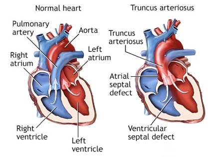
Truncus arteriosus is a rare type of congenital heart disease characterized by a single blood vessel arising from the right and left ventricles, instead of the normal two (pulmonary artery and aorta).
There are four subtypes of truncus arteriosus, depending on the specific anatomy of the single vessel.
Causes
In normal circulation, the pulmonary artery arises from the right ventricle, and the aorta arises from the left ventricle, which are separate from each other. Coronary arteries, which supply blood to the heart muscle, arise from the aorta, just above the valve at the entrance of the aorta.
In truncus arteriosus, a single arterial trunk arises from the ventricles. A large ventricular septal defect (hole between the two ventricles) is usually also present. As a result, the blue (unoxygenated) and red (oxygenated) blood mix completely.
Some of this mixed blood goes to the lungs, some to the coronary arteries, and the rest to the body. Usually, too much blood is sent to the lungs. Meanwhile, the blood going to the coronary arteries and the rest of the body often does not contain enough oxygen.
If left untreated, two problems occur. First, the lungs are filled with fluid, making it difficult to breathe. The second problem is that the blood vessels to the lungs become narrow and are permanently damaged. Over time, it becomes very hard for the heart to force blood to them. This is called pulmonary hypertension and it can be life-threatening.
Truncus arteriosus is very rare.
Symptoms
•Heart failure
•Lethargy
•Poor feeding
•Shortness of breath (dyspnea)
•Rapid breathing (tachypnea)
•Fatigue
•Cyanosis (blue discoloration of skin)
•Delayed growth or growth failure
•Clubbing (broadening) of the finger tips
Exams and Tests
The cardiologist or pediatrician usually hears a murmur when listening to the heart with a stethoscope.
•ECG shows signs of enlargement of the heart (ventricular hypertrophy).
•X-ray of the chest shows heart enlargement and fluid-filled lungs.
•Echocardiogram shows a ventricular septal defect (VSD) and a single truncal artery -- definitive diagnosis.
•Rarely, a heart catheterization is necessary to help with the diagnosis or planning of a treatment strategy.
•MRI of the heart.
Treatment
Surgery is needed to treat this condition. Two procedures are available. One is banding of the pulmonary arteries coming off the truncus, but it is rarely used anymore. The other procedure is called complete repair. Complete repair appears to be the preferred option but as the child grows, repeat surgical procedures may be necessary.
Outlook (Prognosis)
Complete repair usually provides good results. Re-operation may be necessary as the patient grows. Untreated cases have a poor outcome, usually leading to death between during the first year of life. Rarely, the diagnosis is missed until early adulthood; these patients generally need a heart and a lung transplant.
Possible Complications
•Heart failure
•Pulmonary hypertension (high blood pressure in the lungs) with pulmonary obstructive lung disease
When to Contact a Medical Professional
Call your health care provider if your infant or child appears lethargic, does not eat well, appears excessively tired or mildly short of breath, or does not seem to be growing or developing normally.
If the child's skin, lips, or nail beds appear blue or if the child seems significantly short of breath, take the child to the emergency room or have the child examined promptly.
Prevention
There is no known prevention, but early treatment can often prevent serious complications.
Alternative Names
Truncus
References
Zipes DP, Libby P, Bonow RO, Braunwald E, eds. Braunwald's Heart Disease: A Textbook of Cardiovascular Medicine, 8th ed. St. Louis, Mo; WB Saunders; 2007.

Truncus Arteriosus